Article

닭 배아 가장자리 상피조직 융합 과정의 분자 및 세포적 특성

정이진1https://orcid.org/0009-0004-6857-9589, 이형철2,https://orcid.org/0000-0001-5438-1900
Yi Jin Jung1https://orcid.org/0009-0004-6857-9589, Hyung Chul Lee2,https://orcid.org/0000-0001-5438-1900
Author Information & Copyright
1전남대학교 생명과학기술학부 학부생
2전남대학교 생명과학기술학부 교수
1Student, School of Biological Sciences and Technology, Chonnam National University, Gwangju 61186, Republic of Korea
2Professor, School of Biological Sciences and Technology, Chonnam National University, Gwangju 61186, Republic of Korea
To whom correspondence should be addressed : hyungchul@jnu.ac.kr

© Copyright 2024, Korean Society of Poultry Science. This is an Open-Access article distributed under the terms of the Creative Commons Attribution Non-Commercial License (http://creativecommons.org/licenses/by-nc/4.0/) which permits unrestricted non-commercial use, distribution, and reproduction in any medium, provided the original work is properly cited.

Received: Nov 02, 2024; Revised: Nov 10, 2024; Accepted: Nov 19, 2024

Published Online: Dec 31, 2024

적 요

초기 발달과정 동안 닭 배아는 난황을 따라 확장(외적, epiboly)하며 4일 정도에 난황 전체를 감싸게 된다. 이 배아의 확장은 blastoderm의 가장 바깥 쪽에 존재하는 가장자리세포에 의해 주도된다. 초기 확장 단계와 다르게 epiboly의 마지막 단계, 즉 상피조직이 만나 닫히는 과정은 기술적 어려움으로 연구가 되어있지 않다. 본 연구에선 그 상피조직 융합 단계의 간접적인 연구를 위해 초기 배아를 반으로 잘라 마주보게 한 뒤 ex-ovo culture를 통해 분석하였다. 두 상피조직 판이 만나 접합하는 과정동안 가장자리세포 특이적 마커인 DACT2, RNH1SNAI2 발현 양상이 변화하는 것이 관찰되었다. 세 마커 모두 상처 가장자리에서도 발현을 보였으며 이는 발생과정의 조직융합과 상처치유과정에서 유사한 분자세포학적 원리가 공유될 수 있음을 시사한다. 또한, 상피조직 융합과정 동안 세포의 이동 및 섞임 현상 관찰을 위해 한쪽 배아를 DiI로 염색한 뒤 실험을 수행하였으며, 오랜 시간이 지나도 대대적인 세포이동은 관찰되지 않았다. 본 연구 결과는 여러가지 가장자리 조직에서 상황에 따라 분자 및 세포적 특징에 변화가 있음을 보여준다.

ABSTRACT

During the first 4 days of incubation, the chick blastoderm expands and surrounds the yolk by epibolic movement. This expansion is driven by ‘edge cells’, located at the most margin of the embryo. Unlike the expansion, how edge cells behave when the opposing ends of the embryo meet at day 4 and whether there are changes in gene expression during the tissue fusion have not been explored. A major obstacle to studying the final stages of chick epiboly is the technical difficulty in monitoring embryos at those developmental stages over time. In this study, we mimic the final stage of the epiboly by confronting the two isolated embryonic halves from an early embryo during ex-ovo culture. The results show changes in the expression of the markers that are known to be expressed strongly in the edge cells, such as DACT2, RNH1, and SNAI2, upon the tissue fusion. All the markers are also newly expressed in the wound edges made by cutting the embryo in half, suggesting a possibility of similar cellular mechanisms between developmental tissue fusion and wound healing process. Also, to investigate cell migration across the border between the opposing tissue after fusion, one of the isolated embryonic halves is stained with a lipophilic dye, DiI. As a result, no prominent cell migration from one side to the other side is observed. Our results show distinct molecular and cellular characteristics in tissue edges of different situations.

Keywords: 닭; 가장자리세포; 외적; 조직융합; 상처치유
Keywords: chick; edge cells; epiboly; tissue fusion; wound healing

서 론

배아 발생 과정의 여러 단계에서 마주보며 다가오는 두 상피조직 판이 접촉하고 융합되어 하나의 연속적인 구조를 형성하는 경우가 다수 존재한다(Keller, 2002; Ray and Niswander, 2012). 이러한 조직융합 과정은 척추동물의 배아 발달 중 여러 장기 및 조직의 형성의 기본 원리 중 하나이며, 이 과정에서의 문제는 선천성 기형으로 이어질 수 있다(Perez-Pomares and Foty, 2006; Ray and Niswander, 2012). 또한 일반적 발생과정뿐 아니라, 표피가 손상된 곳에서 스스로 조직을 복구하는 상처치유과정에서도 관찰된다(Jacinto et al., 2001). 상피조직 판의 움직임은 주로 가장자리에 위치한 세포의 이동에 의해 주도되며(Petrie and Yamada, 2012), 이는 세포의 이동하는 방향면의 세포 골격 재편성 및 층상위족 활동에 의한 물리적 장력조절에 의해 제어된다(Onochie et al., 2019). 초기 접합부는 신경관(neural tube) 형성 과정 연구에서 많이 밝혀진 것처럼 상피 시트 조직 판의 앞쪽 가장자리를 따라 형성되어 두 상피를 결합한다(Pyrgaki et al., 2010; Rolo et al., 2016; Vijayraghavan and Davidson, 2017). 새로운 세포 간 접합 생성을 위한 접촉 및 신호 전달을 감지하는 것은 다양한 세포학적 과정(예: actin polymerization)을 포함한 복잡한 현상이며(Vasioukhin et al., 2000), 다른 시스템에서 이러한 현상이 어떻게 발생하는지, 이에 대한 생물학적 원리를 파악하고, 제어하는 방법을 발굴하는 것은 조직 공학 기술의 목표와 밀접하게 연계된다. 그럼에도 불구하고 척추동물에서의 조직융합의 세포 수준의 기작은 아직까지 잘 밝혀져 있지 않고 연구모델 또한 부족하다.

닭 배아는 포유류와 유사한 발생 과정을 가지며, 연구모델로서 실험적, 경제적 접근성이 높아 인간을 포함한 고등 척추동물의 발생 메커니즘을 연구하는 데 유용하다. 닭 배아는 발달 단계가 명확히 구분되며(Hamburger and Hamilton, 1951), 여러 발생단계에 세포 및 조직의 변화를 비교적 쉽게 추적할 수 있다. 원반형태의 닭 배아는 부화 과정동안 점점 확장하며 4~5일이 지나면 난황 전체를 둘러 감싸게 된다. 이 확장은 배아의 가장자리에 위치한 특수한 세포에 의해 주도되며, 이 ‘가장자리’ 세포는 배아 내 존재하는 모든 세포 중에 난황막의 안쪽(난황을 향한) 표면에 부착되는 유일한 세포군이다(New, 1959; Lee et al., 2022). 일반적인 상피 조직의 융합 또는 상처치유과정에서의 인접한 상피 조직의 앞쪽 가장자리에선 세포 위족이 형성되며 조직융합을 주도한다고 알려져 있다(Martin and Parkhurst, 2004). 이와 유사하게, 넓고 평평한 층상위족 및 뾰족한 사상위족이 닭 배아의 가장자리 세포에서 뻗어 나오며, 이를 통해 닭 배아 확장과정 동안 가장자리 세포는 다른 상피조직융합 과정과 유사한 방식으로 이동하는 것으로 생각된다(Bellairs, 1963; Bellairs et al., 1969; Lee et al., 2022).

그동안 시험관 및 생체 내 상처 치유 연구에서, 초파리와 같이 유전적으로 다루기 쉬운 종의 형태발생적 움직임에 대한 분석을 통해 상피 융합에 관한 신호와 세포운동성을 유발하는 세포골격을 식별한 바 있다(Affolter et al., 2009; Polyakov et al., 2014). 하지만 척추동물의 초기배아의 상피조직에 대한 연구는 초기 발생단계 배아 회수의 어려움, 체외배양 기술의 한계 등으로 인해 많이 알려져 있지 않다. 본 연구에서는 선행 연구에서 밝혀진 닭 배아의 가장자리 세포의 특성과 상피세포 융합 과정의 유사점(Lee et al., 2022)을 이용해 닭 배아에서의 상피 융합 과정을 관찰하며 특히 닭 배아의 epiboly의 마지막 단계를 간접적으로 관찰한다. 또한 가장자리세포에서 특이적으로 발현하는 유전자 마커를 활용하여 상피조직융합 과정동안 유전자별 발현 변화를 분석한다. 마지막으로 조직융합 후 세포의 추가이동 및 섞임 여부 검증을 위해 세포 염색을 통해 관찰한다.

재료 및 방법

1. 배아 배양 및 체외 배양

수정된 하이라인 브라운(Hy-line Brown) 종 계란(대한민국 나주, 벧엘농장)을 38°C에서 24시간 동안 배양하여 H&H stage 6—7인 배아를 획득하였다(Hamburger and Hamilton, 1951). 획득한 배아는 난의 내부와 유사한 환경에서 미세수술 및 실험적 조작을 하기 위해 New가 개발한 체외배양법을 통해 다음과 같이 배양시켰다(New, 1955).

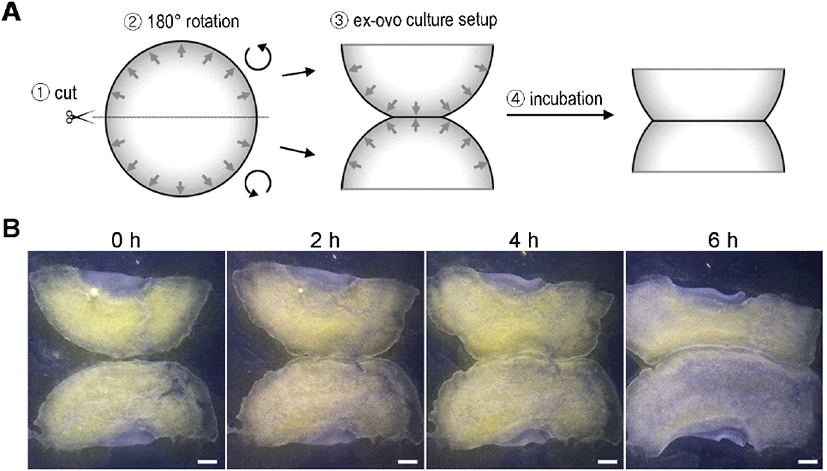
난각의 반(첨단부 또는 둔단부)을 제거한 뒤, 약숟가락을 사용하여 알부민을 최대한 제거한다. 이때, 체외배양 시 필요한 묽은 알부민은 모아둔다. 알부민을 제거한 달걀을 Pannett-Compton 수용액(Pannett and Compton, 1924)에 담근 채, 난황을 둘러싼 난황막을 벗겨내고 배아를 난황막에서 분리한다. 난황막은 링에 씌우고 배아는 반으로 잘라 배아의 가장자리 부분이 서로 마주 보도록 하여(배아의 반쪽만 180° 회전) 난황막 위에 올려준다(Fig. 1A). 이때, 난황막과 배아가 모두 난황 쪽을 향했던 부분이 위로 가도록 하며 방향을 일치시킨다. 물기를 최대한 제거하여 막에 배아를 밀착시킨 후, 2.5 mL 묽은 알부민이 담긴 35 mm 배양접시에 링 째로 막과 배아를 옮겨준다. 이것을 38°C 배양기에서 5~6시간 동안 키워준다.

kjps-51-4-251-g1
Fig 1. Epithelial tissue fusion of the edge of the early chick embryo over time. (A) Schematic diagram of experimental procedure. When an HH stage 6 embryo is cultured ex-ovo with its edges facing each other after being cut in half, the opposing edges attach gradually over time. (B) Representative bright-field images at different time points during epithelial tissue fusion (n>3). About 1/3 edge region is attached after 2 hours of incubation and about 2/3 edge region is attached after 6 hours. Scale bars: 1 mm.
Download Original Figure

목적한 시간만큼 배양된 배아는 고정을 위해 4% paraformaldehyde를 적당량 처리한 후, 날카로운 포셉으로 난황막으로부터 분리한다. 막에서 분리된 배아를 4% paraformaldehyde에 보관한 채 4°C에서 24시간 또는 상온에서 1시간 고정시킨 후, 최소 24시간 동안 100% 메탄올에서 탈수시킨다.

2. Whole-Mount mRNA In Situ Hybridization

고정시킨 배아는 기존에 알려진 mRNA in situ hybridization을 거쳐 유전자 발현을 확인한다(Stern, 1998). 먼저, 차례대로 75%, 50%, 25% 메탄올에서의 수화 과정을 거친 후, 효과적인 프로브 및 항체 침투를 위해 proteinase K 처리를 통해 처리한다. 0.1% glutaraldehyde가 포함된 4% paraformaldehyde에서 30분 동안 후고정시킨다. 이후, 발현을 확인할 유전자의 프로브를 사용하여 70°C에서 hybridization 과정을 하루 동안 수행한다. 본 실험에서 사용한 유전자는 dishevelled binding antagonist of beta catenin 2(DACT2), ribonuclease/angiogenin inhibitor 1(RNH1), 그리고 snail family transcriptional repressor 2(SNAI2)이다(Lee et al., 2022). 그 후, 여러 번의 세척 과정을 거친 후, blocking buffer(1 mg/mL의 bovine serum albumin과 5%의 heat-inactivated goat serum이 포함)에서 3시간 배양시킨다. Anti-Digoxigenin AP antibody(11093274910, Sigma-Aldrich)를 blocking buffer에 희석(1:5,000)시킨 용액으로 교체한 뒤, 4°C에서 하루 동안 배양해 항체를 부착시킨다. 그 후, antibody 세척 과정을 거친 후 NBT-BCIP를 포함된 발색 용액을 배아에 처리해 발현을 관찰한다. 발색이 완료된 배아는 4% paraformaldehyde에서 장기간 보관할 수 있다.

3. Lipophilic Tracer, DiI(1,1‘-Dioctadecyl-3,3,3’,3‘-tetramethylindocarbocyanine Perchlorate) 염색

2 mM DiI(C7000, ThermoFisher) 1 μL을 Pannett-Compton 수용액 500 μL와 혼합해 염색 용액을 만든다. 반으로 자른 배아의 한 쪽은 염색 용액에 담그고, 나머지 한 쪽은 Pannett-Compton 수용액에 담근 후, 38°C 배양기에서 30분간 배양해 배아를 염색시킨다. 염색 후 체외배양 전에 Pannett-Compton 수용액에서 수 차례 세척한다.

4. 현미경 관찰

DiI 염색을 거친 샘플을 제외한 모든 배아는 실체 현미경(LEICA S9D) 및 광학삼안현미경(MIC S16C)을 사용하여 관찰 및 촬영하였다. DiI 염색된 배아는 형광현미경(LEICA DM IL LED, CoolLED pE-300)으로 관찰 및 촬영하였다.

5. 이미지 분석 및 통계분석

유전자 발현 두께 정량은 ImageJ(Schneider et al., 2012)를 사용하였다. 측정값에 대한 평균 도표화는 PRISM 6 프로그램을 사용하였다. 동 프로그램을 사용하여 One way ANOVA 분석을 통해 평균비교를 한 뒤 Tukey’s 다중비교를 사용해 사후검정을 하였다.

결과 및 고찰

1. 배아 가장자리의 상피조직 접합 관찰

닭 배아의 확장과정(epiboly) 동안 가장자리 조직(상피조직)은 난황을 따라 감싸며 4일 정도 후에 전체 난황을 덮게 된다. 하지만 얇은 세포층으로만 이루어진 그 단계의 특성상 조직 회수가 어렵고 시간별 관찰이 쉽지 않아 연구가 되어 있지 않다. 본 연구에선 닭 배아의 epiboly의 마지막 단계, 즉 조직이 닫히는 과정을 간접적으로 관찰하기 위해 원반형태의 배아를 반으로 잘라 서로 마주보게 한 뒤 체외배양을 수행하며 배아의 모습을 촬영했다(Fig. 1A). 배양 시간이 지남에 따라 배아가 확장하면서 서로 마주보는 가장자리 조직끼리 점진적으로 붙는 모습이 관찰되었다(Fig. 1B). 배양 후 2시간이 경과했을 때는 가장자리 부분의 1/3, 4시간이 지났을 때는 절반 정도가 붙었으며, 6시간이 지났을 때는 가장자리의 2/3정도의 부분이 접합된 모습을 보였다(Fig. 1B). 서로 가까이 위치한 조직부터 먼 조직까지 점진적으로 접합되는 것을 볼 때, 가장자리의 부위별 이동속도는 차이가 없는 것을 생각된다. 또한 반으로 자른 상처부위의 경우, 난황막에 부착 및 조직확장이 되지 않는 것으로 보아, 부착 및 확장하는 특성은 원래 배아의 가장자리세포에 국한되며 대부분의 지역에서 새로 획득되지 않는 것으로 확인된다(Fig. 1B) (New, 1959; Lee et al., 2022).

2. 조직접합 및 난황막 부착에 따른 유전자 발현 변화

다음으로 가장자리 조직의 융합 여부 및 시간 및 난황막 부착 여부에 따른 가장자리 특이적 마커의 발현양상을 확인하기 위해 whole-mount mRNA in situ hybridization을 진행하였다. 마커로는 선행 연구에서 가장자리 세포에서 발현한다고 보고된 DACT2, RNH1, 그리고 SNAI2를 사용하였다(Lee et al., 2022). 세 유전자 모두 상처치유과정에서 특이적으로 발현할 뿐 아니라 치유과정을 매개하는 중요한 역할을 하는 것으로 알려져 있다(Hudson et al., 2009; Kim et al., 2020; Neu et al., 2024). 마커의 지역별 발현비교를 위해, 배아에서 총 4가지 지역(I-IV)을 다음과 같이 구분하였다(Fig. 2A): (I) 접합된 후 시간이 경과된 가장자리(closed edge, old), (II) 접합된 지 얼마 안 된 가장자리(closed edge, young), (III) 접합되지 않은 가장자리(free edge), (IV) 배아를 반으로 잘랐을 때의 상처부위(cut edge, wound). 또한 각 지역에서의 발현차이를 구체적으로 분석하기 위해 발현 부위의 두께를 측정하여 정량 및 비교하였다. 평균 비교를 위해 사용된 유전자별, 배아의 수는 최소 5, 총 측정지역의 수(Figs. 2H, 2I, and 2J의 n수)는 최소 15 이상이다.

kjps-51-4-251-g2
Fig 2. Expression patterns of the edge-cell-markers, DACT2, RNH1, and SNAI2, checked by in situ hybridization during epithelial tissue fusion of the edge of the early chick embryo. (A) Schematic diagram of experimental procedure. The four different monitored regions after the procedure are marked in red boxes: old closed edge, I; young closed edge, II; free edge, III, cut edge (wound), IV. Expression patterns of DACT2 (B, E), RNH1 (C, F), and SNAI2 (D, G) (n>5 for each gene). (E—G) Magnified views of the marked regions in red boxes in (B—D) of each gene. (H—J) Quantification of the thickness of the expression of each gene in the four different regions. Black dots and blue lines indicate mean and standard error of mean, respectively. Values with different letters are significantly different (P<0.05, oneway ANOVA with Turkey’s multiple comparisons). Scale bars: 1 mm in (B—D), 200 μm in (E—G).
Download Original Figure

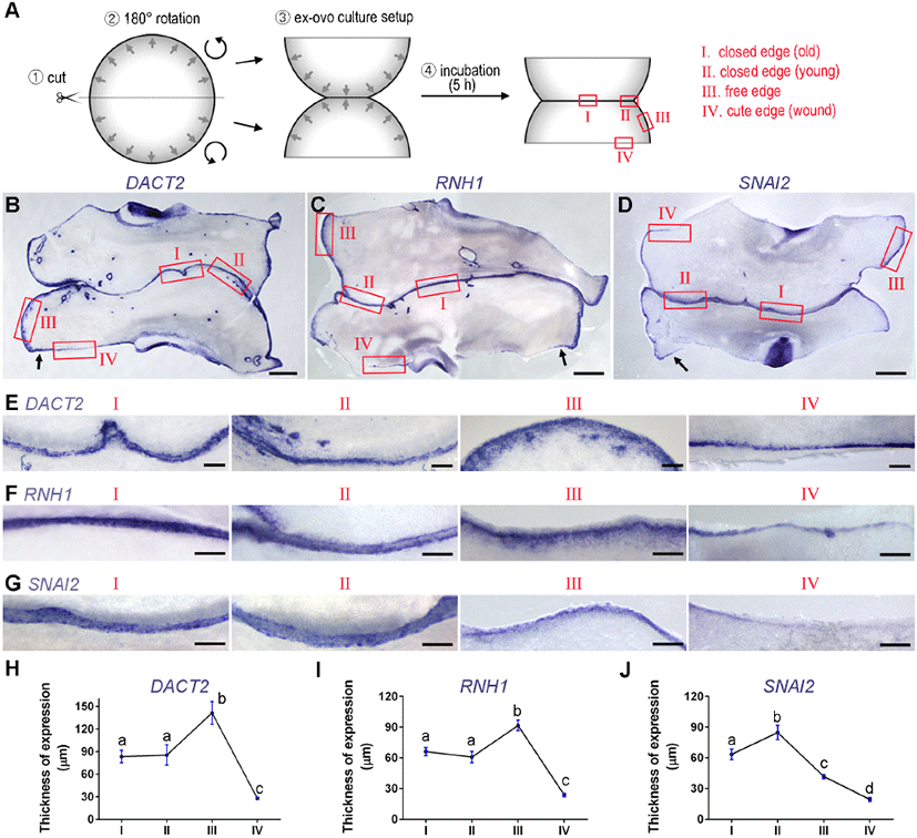
세 유전자 모두, 배아 내 발현(expression in the embryonic tissue)을 제외하고 바깥의 배아외조직(extraembryonic tissue)의 가장자리에서 특이적인 발현을 보였으며 다음과 같은 결과를 보였다. 먼저, 세 유전자 모두 free edge인 III 지역에서 정상 배아의 가장자리보다 두꺼운 발현(최소 30 μm~최대 100 μm 이상)을 보였다 (Figs. 2 B, 2C, 2D, 2E, 2F, and 2G의 III 지역들). 이는 기존에 보고된 바와 같이, 준비과정에서 배아가 막에서 분리된 후 재부착(reattachment)하는 과정을 통해 생긴 현상으로 보인다(Lee et al., 2022). 발현 두께의 증가는 가장 바깥쪽(가장자리)보다 안쪽(비가장자리)에 위치한 세포 들에서의 신규발현을 의미하며, 이는 reattachment 과정 중 비가장자리 세포들을 포함한 배아의 바깥쪽에 있는 세포들이 가장자리세포화(가장자리세포의 특성을 획득)되는 것을 시사한다(New, 1959). 즉, reattachment를 통한 물리적 환경 변화가 배아 끝 쪽의 세포를 언제든 붙을 수 있는 상태로 준비시키는 것으로 생각된다.

두 번째로 세 유전자에서 조금 다른 형태로 접합 후에 발현 두께가 줄어드는 것이 관찰되었다. DACT2와 RNH1의 경우, free edge(III)에 비해 closed edge(I 및 II)에서 두께가 줄어들었고(Figs. 2B, 2C, 2E, 2F, 2H, and 2I), SNAI2의 경우 young closed edge(II)에 비해 old closed edge(I)의 발현 두께가 줄어 들었다(Figs. 2D, 2G, and 2J). 두 경우의 형태는 다르지만, 발현 두께가 줄어드는 것은 가장자리 조직의 접합에 의해 free edge가 없어지면서, 더 이상 비가장자리 세포들의 가장자리세포화가 필요 없어진 것으로 사료된다. 조직 접합 후에 가장자리 조직의 막 부착 상태의 변화에 따른 원인인지는 확인되지 않았다.

마지막으로 배아 가장자리뿐 아니라 cute edge(IV), 즉 상처부위에도 세 유전자의 발현이 관찰 되었다(Figs. 2B, 2C, 2D, 2E, 2F, and 2G의 IV지역들). 다른 나머지 지역(I—III)에 비해, 1—3 세포층 두께의 얇은 발현 양상이 관찰되었다(Figs. 2H, 2I, and 2J). 이는 세 유전자가 다양한 상황의 가장자리에 위치한 세포에서 공통적으로 발현함을 보여주며, 일반적인 gap closure와 wound closure에서의 가장자리세포가 유사한 원리로 closure를 주도할 것이라는 추측을 가능케 한다. 추가적으로 배아의 가장 바깥쪽으로부터 500~1,000 μm 거리 안에 있는 cut edge(IV)의 경우, 얇은 발현을 보이는 다른 IV 지역과 달리 free edge(III)와 같은 두께의 발현을 보였다(arrows in Figs. 2B, 2C, and 2D). 이 결과는 두 가지 원인을 추측할 수 있는데, 첫 번째는 주변 가장자리세포의 이동으로 인한 발현증가, 두 번째는 비가장자리세포의 가장자리세포화이다. 기존 연구에서 배아 바깥 쪽에 가까운 비가장자리세포의 경우 wound 상황에서 가장자리세포처럼 막에 부착 및 조직확장을 보이는 것으로 보아 두 번째 설명이 더 가능성이 높은 것으로 생각된다(New, 1959).

3. 상피세포 융합 과정에서 세포의 이동

다음으로 가장자리 조직의 접합 과정에서 마주보는 조직의 세포의 섞임 및 이동 여부를 관찰하기 위한 실험을 진행하였다. 배아를 반으로 잘라 가장자리 부분끼리 맞붙여 배양하는 것은 동일하지만, 반으로 자른 배아에서 한 쪽만 세포막 및 일부 세포질을 염색하는 지질친화성 염색약인 DiI로 처리한 뒤 조직접합 및 배양을 진행하였다(Fig. 3A, n>10). 배양 전과 배양 후 16시간의 배아를 각각 형광 현미경으로 관찰한 뒤 middle과 hinge 부분의 세포 이동양상을 분석하였다(Figs. 2A and 2B).

kjps-51-4-251-g3
Fig 3. Cell mixing during epithelial tissue fusion of the edge of the early chick embryo. (A) Schematic diagram of experimental procedure. One of the isolated half-embryo is stained with a lipophilic dye, DiI, then is confronted with the other half, followed by 16 h of incubation. Both the middle and hinge regions of the fused area are investigated for cell mixing. (B) Bright-filed and fluorescent (DiI) images of edge regions right after ex-ovo culture setup (0 h) and 16 h of incubation. The arrow in the bottom left panel indicates the migration of DiI-positive cells towards the opposing isolated half-embryo. Scale bars: 500 μm.
Download Original Figure

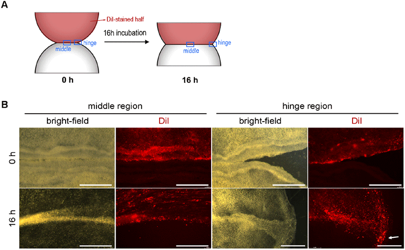
16시간 배양 후 형광 현미경으로 관찰한 결과, middle 지역에서는 마주보는 조직끼리 붙은 경계를 기준으로 (소수의 세포를 제외하고) 이동한 세포가 거의 관찰되지 않았다(Fig. 3B). 하지만, hinge 지역, 즉 가장자리 조직끼리 접합이 시작되는 부분에서는, DiI 염색된 배아의 세포들의 다수가 반대 반쪽 배아 쪽으로 이동한 것이 관찰되었다(Fig. 3B). 이러한 배아의 경우 hinge 부분이 움푹 들어간 곳 없이 매끄럽게 메꿔진 것을 보아, 접합된 경계를 따라 이동한 결과라고 예상할 수 있다. 하지만 왜 hinge에 위치한 세포들은 다른 행동을 보이는지에 대한 설명은 추가 연구가 필요하다.

사 사

이 논문은 2024년도 정부(교육부)의 재원으로 한국연구재단의 G-램프(LAMP) 사업의 지원을 받아 수행된 연구임(No. RS-2024-00442775).

REFERENCES

1.

Affolter M, Zeller R, Caussinus E 2009 Tissue remodelling through branching morphogenesis. Nat Rev Mol Cell Biol 10(12):831-842.

2.

Bellairs R 1963 Differentiation of the yolk sac of the chick studied by electron microscopy. Development 11(1):201-225.

3.

Bellairs R, Boyde A, Heaysman JE 1969 The relationship between the edge of the chick blastoderm and the vitelline membrane. Wilhelm Roux Arch Entwickl Mech Org 163(2):113-121.

4.

Hamburger V, Hamilton HL 1951 A series of normal stages in the development of the chick embryo. J Morphol 88(1):49-92.

5.

Hudson LG, Newkirk KM, Chandler HL, Choi C, Fossey SL, Parent AE, Kusewitt DF 2009 Cutaneous wound reepithelialization is compromised in mice lacking functional Slug (Snai2). J Dermatol Sci 56(1):19-26.

6.

Jacinto A, Martinez-Arias A, Martin P 2001 Mechanisms of epithelial fusion and repair. Nat Cell Biol 3(5):E117-E123.

7.

Keller R 2002 Shaping the vertebrate body plan by polarized embryonic cell movements. Science 298(5600):1950-1954.

8.

Kim DH, Kim EJ, Kim DH, Park SW 2020 Dact2 is involved in the regulation of epithelial-mesenchymal transition. Biochem Biophys Res Commun 524(1):190-197.

9.

Lee HC, Fadaili Y, Stern CD 2022 Molecular characteristics of the edge cells responsible for expansion of the chick embryo on the vitelline membrane. Open Biol 12(9):220147.

10.

Martin P, Parkhurst SM 2004 Parallels between tissue repair and embryo morphogenesis. Development 131(13):3021-3034.

11.

Neu C, Beckers C, Frank N, Thomas K, Bartneck M, Simon TP, Mossanen J, Peters K, Singendonk T, Martin L, Marx G, Kraemer S, Zechendorf E 2024 Ribonuclease inhibitor 1 emerges as a potential biomarker and modulates inflammation and iron homeostasis in sepsis. Sci Rep 14(1):14972.

12.

New DA 1959 The adhesive properties and expansion of the chick blastoderm. J Embryol Exp Morphol 7:146-164.

13.

New DAT 1955 A new technique for the cultivation of the chick embryo in vitro. J Embryol Exp Morph 3:326-331.

14.

Onochie OE, Zollinger A, Rich CB, Smith M, Trinkaus-Randall V 2019 Epithelial cells exert differential traction stress in response to substrate stiffness. Exp Eye Res 181:25-37.

15.

Pannett CA, Compton A 1924 The cultivation of tissues in saline embryonic juice. Lancet 1:381-384.

16.

Perez-Pomares JM, Foty RA 2006 Tissue fusion and cell sorting in embryonic development and disease: biomedical implications. Bioessays 28(8):809-821.

17.

Petrie RJ, Yamada KM 2012 At the leading edge of three-dimensional cell migration. J Cell Sci 125(Pt 24):5917-5926.

18.

Polyakov O, He B, Swan M, Shaevitz JW, Kaschube M, Wieschaus E 2014 Passive mechanical forces control cell-shape change during Drosophila ventral furrow formation. Biophys J 107(4):998-1010.

19.

Pyrgaki C, Trainor P, Hadjantonakis AK, Niswander L 2010 Dynamic imaging of mammalian neural tube closure. Dev Biol 344(2):941-947.

20.

Ray HJ, Niswander L 2012 Mechanisms of tissue fusion during development. Development 139(10):1701-1711.

21.

Rolo A, Savery D, Escuin S, de Castro SC, Armer HE, Munro PM, Mole MA, Greene ND, Copp AJ 2016 Regulation of cell protrusions by small GTPases during fusion of the neural folds. Elife 5:e13273.

22.

Schneider CA, Rasband WS, Eliceiri KW 2012 NIH Image to ImageJ: 25 years of image analysis. Nat Methods 9(7):671-675.

23.

Stern CD 1998 Detection of multiple gene products simultaneously by in situ hybridization and immunohistochemistry in whole mounts of avian embryos. Curr Top Dev Biol 36:223-243.

24.

Vasioukhin V, Bauer C, Yin M, Fuchs E 2000 Directed actin polymerization is the driving force for epithelial cell-cell adhesion. Cell 100(2):209-219.

25.

Vijayraghavan DS, Davidson LA 2017 Mechanics of neurulation: From classical to current perspectives on the physical mechanics that shape, fold, and form the neural tube. Birth Defects Res 109(2):153-168.